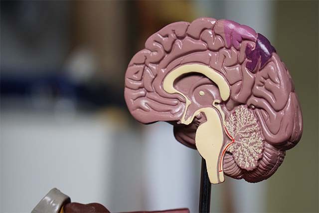
Applying deep brain stimulation to a different region in the brain than has been used for other conditions improved the recovery of lower limb movements in two patients with severe spinal cord injuries, researchers reported.
Deep brain stimulation applied to the lateral hypothalamus “immediately augmented walking” in mice and rats and in the two humans, according to the report published in Nature Medicine.
This type of stimulation has been used to treat Parkinson’s disease and other movement disorders, targeting other brain regions, but had not been tried for spinal injuries.
In both patients, while the spinal cord was damaged it still had the ability to send some signals to or from the brain.
“Once the electrode was in place and we performed the stimulation, the first patient immediately said, ‘I feel my legs.’ When we increased the stimulation, she said, ‘I feel the urge to walk!’
“This real-time feedback confirmed we had targeted the correct region, even if this region had never been associated with the control of the legs in humans,” study leader Jocelyne Bloch of Ecole Polytechnique Federale de Lausanne said in a statement.
“At this moment, I knew that we were witnessing an important discovery.”
The other patient, a 54-year-old who had been in a wheelchair since a 2006 ski accident, said that soon after the treatment, he was able to walk “a couple of steps” and “reach things in my cupboards in the kitchen.”
Both patients also had long-term improvements that persisted even when the stimulation was turned off, the researchers said.
Knee arthritis eased by blocking of arteries
A minimally invasive procedure can provide significant relief from knee osteoarthritis and may prevent the need for knee replacement surgery, according to data being presented this week at the Radiological Society of North America meeting in Chicago.
The 403 patients in the study, ages 40 to 90, underwent genicular artery blocking, or embolization, to treat moderate to severe knee osteoarthritis. The genicular arteries surround the knee joint.
In osteoarthritis, abnormal blood vessels can branch out from these arteries, grow into the bone, become inflamed or compressed, and cause pain, swelling and loss of function and mobility. Blocking the abnormal vessels helps disrupt those effects, researchers found.
A year after genicular artery embolization, patients’ scores on measures of quality-of-life and pain had improved by 87% and 71%, respectively, the researchers reported.
The treatments were particularly effective in patients with early-stage knee osteoarthritis, they said.
The procedure “can effectively reduce knee pain and improve quality of life early after the treatment, with these benefits being maintained over the long term,” study leader Dr. Florian Nima Fleckenstein of Charite University Hospital Berlin said in a statement.
This was especially true for people who have not had success with treatments like physical therapy or pain medications, Fleckenstein added.
“This could potentially offer a new lease on life for many patients who suffer from debilitating pain and mobility issues caused by osteoarthritis.”
New type of ink could replace electrodes for brain monitoring
Researchers have designed a new type of ink that can be painted onto a patient’s scalp to allow measurement of brain activity, potentially replacing electrodes with long wires.
The current method for measuring brain activity – electroencephalography (EEG) – requires technicians to measure the patient’s scalp with rulers and pencils and mark over a dozen spots where they will glue on the electrodes connected to a machine.
“Our study can potentially revolutionize the way non-invasive brain-computer interface devices are designed,” Jose Millan of the University of Texas at Austin, who worked on the project, said in a statement.
With a computer algorithm, researchers identify the spots on the scalp for measuring brain activity. They used a digitally controlled inkjet printer to spray a thin layer of the ink onto those spots. Once dried, the ink works as a thin-film sensor, picking up brain activity through the scalp.
Electronic tattoos have already been used on hairless skin for measuring heart activities, but designing e-tattoos for hairy skin has been a persistent challenge, the researchers noted in a report published in Cell Biomaterials.
To overcome this, they designed an ink that can flow through hair to reach the scalp.
When the team printed e-tattoo electrodes onto the scalps of five short-haired volunteers and attached conventional electrodes next to the e-tattoos, the e-tattoos detected brainwaves as efficiently as the electrodes.
The e-tattoo electrodes showed stable connectivity for at least 24 hours, while gel used on the conventional electrodes started to dry out after six hours. Beyond six hours, over a third of the conventional electrodes were failing to pick up any signal, and most of the others had reduced contact with the skin, resulting in less accurate signal detection.
“The broader significance of this technology lies in its potential applications beyond traditional EEG use,” the researchers said in their report.
“On-scalp printed ultrathin e-tattoos could play a pivotal role in developing brain-computer interfaces for… prosthetics, virtual reality, and human-robot teaming,” they said.
—Reporting by Nancy Lapid; editing by Bill Berkrot

